BenchTop™ Vibration Control Enables Critical Improvements in Microscopy
High-resolution microscopy offers researchers and clinicians a powerful analytical tool to help uncover a variety of biological phenomena and processes. However, instruments of this type require a stable environment in order to function at their peak of accuracy and precision. With the ever-increasing demand to prepare, examine, and then record with more accuracy and precision (via photo or electron-micrography) specimens to study micron and sub-micron structural features, vibration isolation of an experimental set up becomes all the more necessary.
The challenge of designing an appropriate vibration isolation system is formidable because a good vibration isolation system has to provide high-performance resistance to vibrations (which usually requires large free standing isolators), yet still keep a compact low profile for it's users in order to minimize space requirements and maximize equipment accessibility. Newport accomplished both with BenchTop™.
How Does this New Technology Benefit the Life-Science Researcher?
Refer to Figure 1. High-performance BenchTop™ Compact Vibration Isolation Platforms enhance the resolution and accuracy of photomicroscopes, balances and other table top analytical instruments by isolating the work surface from horizontal and vertical vibrations. BenchTop is especially useful for improving the quality of time-exposure photomicrography,which is extremely sensitive to the effects of vibration. The BenchTop unit is a support structure, with either a white plastic-laminate or stainless steel top, and four recessed compact isolators in the platform’s bottom.
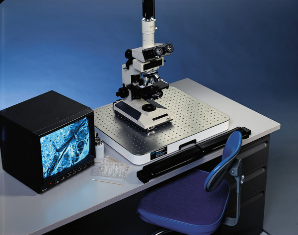
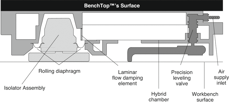
BenchTop units are among the most compact vibration isolation systems available. In sizes: 16 in. x 20 in. (40.6 x 50.8 cm) and 30 in. x 36 in. (76.2 x 91.4 cm), BenchTop incorporates four isolator modules recessed into the bottom of the platform. Both contain a pneumatic flexible rolling diaphragm that supports the load - decoupling vibrations from the isolated platform.
BenchTop’s low profile design adds a mere 2 in. of height above the support surface, allowing comfortable long-term operation using standard chairs or stools. Optional padded armrests, which attach to the edge of the supporting bench or desktop, provide comfortable support during extended microscope or instrument use.
Unique Benefits of Stabilizer™ Technology
- Compact Size
The Stabilizer™ technology in BenchTop™ not only does a better job compared to ordinary vibration isolators, but it uses a much smaller air volume - enabling isolators to be placed underneath the work surface. This eliminates bulky outrigger-style isolators around an experimental platform.
- Faster Settling Times
To avoid work delays after disturbances, short settling times are important to stabilize the isolated microscope or instrument as quickly as possible. Laminar flow damping reduces settling times by about 50% for both large-and-small magnitude disturbances when compared to ordinary isolators.
- Superior High-Center-of-Mass Stability
A critical design consideration in the development of BenchTop™ was the isolation of heavy, higher-center-of-gravity equipment like photomicroscopes used in life science and semiconductor inspection applications.
- Lower Natural Frequency
The lower the natural frequency of the isolator (the “softer” the spring constant), the better the protection against low-frequency vibrations — by far the most critical to instrument performance.
- Patented Self-Centering Piston Mechanism
Automatically self-centers each isolator’s piston for unrestricted vertical travel, guaranteeing the best possible vibration isolation performance.
Experimental Results are the Real Test
Theory is one thing — real results another! Refer to Figures 3 and 4 for just two examples of how this new vibration isolation technology can positively affect the gathering of data.
Figure 3 represents two high magnification photomicrographs (500X) taken of a dendritic spine (process emanating from a mammalian neuron) with (3a) and without (3b) the BenchTop™ compact isolator platform. It is clear that the isolation unit provided the superior image showing fine structural details (dimensions of 1 x 20 microns).


Figure 4 represents a set of electron micrographs of ultra thin tissue sections prepared for the electron microscope before (4a) and after (4b) inflation of the BenchTop™ isolation system. The non-inflated picture (4a) exhibits light and dark bands indicating vibration induced knife chatter during sectioning. These striations are not evident in sections cut from the same tissue block after the BenchTop™ is inflated (4b). NOTE: The dark black lines are the “grid bars” used to support the section.

