Super-Resolution Microscopy
The axial resolution in standard confocal microscopy is limited by the microscope objective and the wavelength of light. Even by using short-wavelength light and an oil immersion objective, the resolution of a standard fluorescence system is limited to around 200 nm. Recently, super-resolution microscopy techniques have been developed which break this limit and allow imaging with an axial resolution down to a few tens of nm. This work was recognized with the 2014 Nobel Prize in Chemistry.
Stimulated Emission Depletion Microscopy (STED)
One super-resolution approach is called stimulated emission depletion (STED) microscopy. This technique uses a confocal microscope and a diffraction limited excitation beam that is scanned across the sample. A second beam (the depletion beam) is spatially shaped like a donut and, when overlapped with the excitation beam, forces the illuminated portion of the sample to emit light at one wavelength. The non-depleted region then emits light at a different wavelength, which is filtered and measured. Because the central hole in the donut can be made much smaller than the diffraction limit, the non-depleted area used for imaging will be smaller as well.
Photoactivatable Localization Microscopy (PALM)
A second approach to obtain super-resolution is classified as localization microscopy. Two of the most popular techniques are called photoactivatable localization microscopy (PALM) and stochastic optical reconstruction microscopy (STORM). A fluorophore is a fluorescent chemical compound that can emit light upon excitation. When measuring the spatial distribution of a single fluorophore through the microscope, the center of the distribution can be determined with greater accuracy than its width. This is the basis for improved resolution using localization microscopy. This approach requires that a small number of fluorophores are emitting in a single captured frame and that there is no overlap with neighboring emitters. In order to make up for the necessarily weak signal strength, the super-resolution image is built up from several hundred thousand images of the localized positions of the individual fluorophores. This can take several minutes and therefore may not be fast enough to capture processes in live cells. The PALM and STORM techniques use slightly different methods to control the behavior of the fluorophores in order to keep the emission well-separated in time.
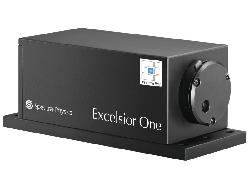
MKS offers a large range of CW and quasi-CW lasers of low to moderate output powers that can be used for super-resolution microscopy and other bioimaging applications. The Spectra-Physics Excelsior¨ family (see Figure) of low-power CW solid-state and semiconductor lasers offers customers a complete range of wavelengths and power levels. DPSS lasers are available at a variety of wavelengths: 473, 505, 515, 532, 542, 561, 594, and 1064 nm. For applications requiring a 488 nm laser, Spectra-Physics offers an Excelsior laser based on highly reliable, externally frequency-doubled diode laser technology. The Excelsior-One laser is the world's smallest 488 nm laser head with a 40% narrower width than other commercially available lasers. In addition to these wavelengths, laser diodes are also available at 375, 405, 440, 642, and 785 nm. Excelsior lasers provide state-of-the-art performance with the smallest footprint in their class. The Spectra-Physics Excelsior product line provides a consistent platform with the same mechanical footprint over a wide range of power levels and wavelengths. This allows for full component interchangeability among laser heads and controllers within a given architecture, requiring no additional adjustment or optimization.

For additional insights into photonics topics like this, download our free MKS Instruments Handbook: Principles & Applications in Photonics Technologies
Request a Handbook